What is the Vestibular System?
The vestibular system is quite small and often forgotten, but is so crucial for our everyday activities!
The Inner Ear
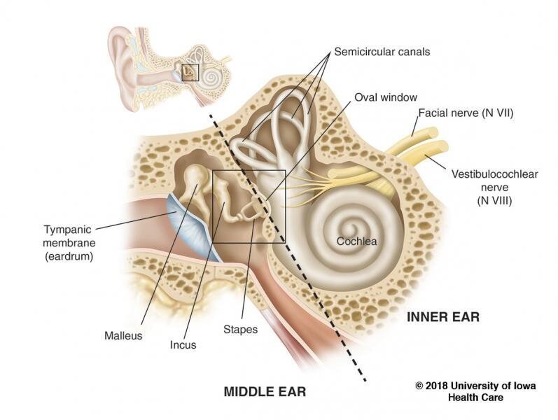
Before we discuss the vestibular system, it is important to note that the vestibular system (as we typically know it) is only one division of the inner ear. The inner ear, which is also known as the labyrinth, is encased in bone called the bony labyrinth, and consists of 2 divisions:
The hearing (auditory) component:
It consists of the cochlea, which is responsible for hearing. Sound waves enter the inner ear and then into the cochlea. The cochlea is filled with a fluid that moves in response to vibrations, which activates nerve endings. These nerve endings transform the vibrations into electrical signals that then travel along the auditory nerve to the brain. The brain interprets these signals as the sounds that we hear.
The balance (vestibular) component:
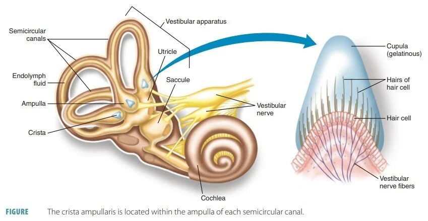
It consists of the peripheral vestibular system, which is responsible for maintaining balance, stability, and spatial orientation. The peripheral vestibular system then sends information to the central vestibular system, which consists of the brain and brainstem. The vestibular system consists of a series of tubes and chambers filled with fluid. The tubes are the semicircular canals (3 in each ear) and the chambers are the utricle and saccule (1 of each in each ear).
The fluid in the inner ear is of a particular composition and there are 2 types of fluid: 1) endolymph is present inside of the semicircular canals and chambers, and 2) perilymph is present outside of the semicircular canals and chambers. Think of the semicircular canals and both types of fluid as “a hose within a hose”. The 2 types of fluid differ in amount and composition and are integral to the proper functioning of the inner ear. Both fluids contain sodium and potassium but in different ratios. There are conditions that affect the stability of these ratios, such as Meniere’s disease, which result in symptoms, such as vertigo, dizziness, nausea, vomiting, ear fullness and tinnitus.
Semi-Circular Canals
In both ears, there are 3 semicircular canals: posterior, anterior, and horizontal. The semicircular canals are located in specific spatial planes. They are oriented roughly 90 degrees to each other (like the corner of a room between two adjoining walls and the floor). Each semicircular canal has one bulbous-shaped end called an ampulla. Within the ampulla is a structure called the cupula, which is a sensory receptor that detects angular movement. The hair cells within the cupula move during angular movement, sending signals to the brain to produce corresponding eye movements.
Otolith Organs
The otolith organs consist of the utricle and saccule and they detect linear acceleration and deceleration, including the linear acceleration of gravity.
The utricle detects linear motion primarily in the horizontal plane (like a car moving forward or backward).
The saccule detects motion primarily in the vertical plane (like an elevator moving up or down).
Each otolithic organ has hair cells embedded in a gelatinous material (like jelly) with calcium carbonate crystals, called otoconia, attached to the surface. The otoconia are sensitive to gravity and allow you to sense movement if there is a change in linear acceleration, even if you may not be moving. They are associated with a very common condition known as Benign Paroxysmal Positional Vertigo, where they dislodge and enter into one of the semicircular canals. As the otoconia are sensitive to gravity, when they enter a semi-circular canal (such as during head movement) that is NOT normally sensitive to gravity, it can result in nystagmus and cause vertigo.
The otolith organs are also associated with neural pathways related to maintaining:
An upright posture of the head and trunk by using the appropriate balance strategies
Gaze stability in response to body tilt or lateral movement
Proper regulation of blood distribution in the body and blood pressure during movement and changes in posture, especially sympathetic nervous system activity (associated with the “fight or flight” response)
The Vestibulocochlear Nerve
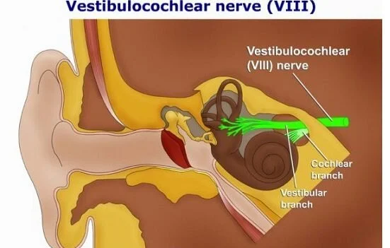
The vestibulocochlear nerve (cranial nerve VIII) travels from the inner ear to the brainstem and is considered part of the peripheral vestibular system until it enters the brainstem. There are 2 portions to the vestibulocochlear nerve: 1) the vestibular nerve is involved with balance, and 2) the cochlear (auditory) nerve is involved with hearing. The vestibular nerve can also be further divided into 2 portions and each portion innervates specific structures of the inner ear: 1) the superior vestibular nerve, and 2) the inferior vestibular nerve.
The vestibular nerves in each ear must work together when sending signals to the brain, so when one nerve’s signals are disrupted, conflicting information is sent to the brain causing vertigo, dizziness, and imbalance. The blood supply follows a similar path as the nerve supply, and when compromised, can result in similar symptoms.
The Oval and Round Windows
The oval and round windows are two openings from the middle ear to the inner ear and are closed off by membranes. They help to transmit sound, but if these structures are compromised, allowing fluid to enter into the middle ear, it can result in symptoms, such as vertigo, imbalance, fullness in the ear, hearing loss, poor tolerance of loud sound, nausea and vomiting. This condition is called a Perilymph Fistula.
Central and peripheral vestibular systems
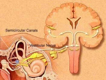
Beyond the peripheral vestibular system, which consists of the inner ear and the pathways to the brainstem, the vestibular system extends into the central nervous system, which consists of the brainstem and brain.
The vestibular primary afferent nerves travel to the vestibular nuclei and the posterior cerebellum. Input from the peripheral vestibular system, as well as input from the visual and somatosensory systems, process and relay:
Neural outputs to control eye movements for gaze stability during movement
Information to the cerebellum to fine tune movements and adapt them based on the task and environment
Information to the brain stem for postural movements, such as equilibrium reactions and postural control when walking
Information to the cerebral cortex for spatial awareness and orientation
Due to the vestibular system’s expansive connections with multiple systems of the body, it is an important sensory organ contributing to multi-sensory integration!


