Vestibular Testing by Audiologists and ENTs: A Guide for Clinicians
A bedside assessment answers most of the questions I need answered to plan vestibular rehabilitation. At the bedside I can't see covert saccades, I can't test the otoliths directly, and a head impulse alone doesn't always tell me whether a loss is unilateral or bilateral. When the answer to one of those questions would change my plan, I refer for formal vestibular testing with an audiologist or otolaryngologist.
Each test covers a different frequency range
The angular vestibulo-ocular reflex (VOR) responds to head movement across a wide frequency range, and each test samples a different part of it. Caloric testing samples very low frequencies. Rotary chair testing covers low to mid frequencies. The video head impulse test (vHIT) measures the high-frequency, high-acceleration range typical of everyday head movement. A patient can be abnormal on one test and normal on another.
The Bárány Society criteria for bilateral vestibulopathy accept any of the three. Along with the typical symptoms, the diagnosis requires bilaterally reduced VOR function on at least one of three measures: horizontal vHIT gain below 0.6 on both sides, a summed peak slow-phase velocity below 6°/s on each side with bithermal calorics, or rotary chair gain below 0.1 at 0.1 Hz (Strupp et al., 2017).
Videonystagmography (VNG)
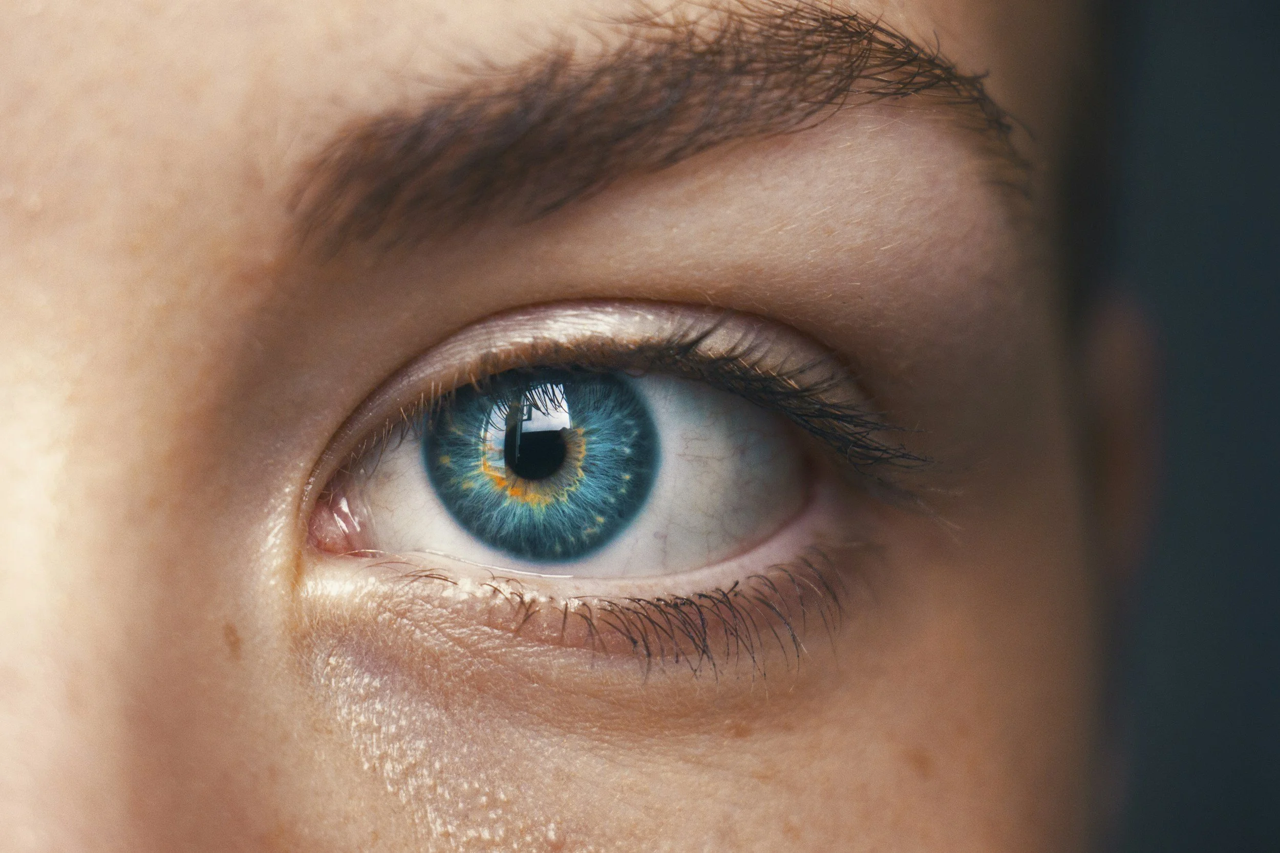
VNG is the video-based version of electronystagmography (ENG). It has three parts.
Oculomotor testing covers saccades, smooth pursuit, optokinetic responses, and gaze holding. Abnormal findings here suggest central involvement, so I check this section before assuming a peripheral loss.
Spontaneous, gaze-evoked, and positional nystagmus are recorded with fixation removed. Peripheral nystagmus is often suppressed by visual fixation and may not be visible at the bedside without Frenzel goggles or video goggles.
Caloric testing irrigates each ear with warm and cold water or air, and the responses are compared to calculate unilateral weakness. Each lab sets its own cut-off for a significant weakness, so I use the threshold printed on the report.
Calorics stimulate one labyrinth at a time without moving the head, and they test a lower frequency range than vHIT. That is why I still find calorics useful when vHIT has already been done.
Video head impulse test (vHIT)
vHIT measures VOR gain for each of the six semicircular canals and records corrective saccades, including covert saccades that happen during the head movement and can't be seen at the bedside. Halmagyi et al. (2017) report that in many dizziness clinics, vHIT has replaced caloric testing as the first test, and sometimes the only test, of canal function.
The same review describes technical sources of error. If the goggles shift on the face during the impulse, the recording shows false eye and head movement. If the goggle sensor is not aligned with the plane of the head movement, the measured head velocity is too low and the calculated gain is falsely high (Halmagyi et al., 2017). When a reported gain doesn't match what I see clinically, I ask the testing clinician about recording quality before changing my plan based on it.
vHIT and calorics can also disagree. McGarvie et al. (2015) reviewed 22 patients with Ménière's disease who had both tests. Responses to caloric stimulation of the affected ear were small or absent, while vHIT gains were in the normal range. The authors proposed that an enlarged, hydropic duct reduces the caloric stimulus without changing the response to rotation. In practice, a normal vHIT in a patient with suspected Ménière's disease does not rule out a caloric weakness.
Vestibular evoked myogenic potentials (VEMPs)
VEMPs are the standard clinical test of otolith function.
The cervical VEMP (cVEMP) is a brief inhibition of the sternocleidomastoid on the same side as the stimulated ear. Air-conducted sound mainly activates the saccule, so the air-conducted cVEMP is used as a test of saccular function.
The ocular VEMP (oVEMP) is a brief excitation of the inferior oblique, recorded under the eye opposite the stimulated ear. It is generated mainly by afferents in the superior vestibular nerve and is used as a test of utricular function.
These attributions are approximate. The superior vestibular nerve also carries a small number of saccular afferents, and bone-conducted stimuli activate the otoliths more generally (Rosengren et al., 2019).
VEMPs are most useful clinically in third window disorders. In superior canal dehiscence, both cVEMPs and oVEMPs show high amplitudes and low thresholds. In vestibular neuritis, a reduced or absent oVEMP with a preserved cVEMP is a common pattern, consistent with the superior division of the nerve being affected more often than the inferior division (Rosengren et al., 2019).
Rosengren et al. (2019) report that the air-conducted cVEMP is absent in about 5 to 15% of healthy adults over 60, and that the air-conducted oVEMP is more often absent and more sensitive to age. They recommend caution before calling an absent air-conducted oVEMP abnormal in an older adult. Methods also vary between labs, so results should be compared with that lab's own normative data.
Rotary chair
Sinusoidal harmonic acceleration testing reports VOR gain, phase, and symmetry across low to mid frequencies. I find it most useful for confirming and grading bilateral loss, for following compensation over time, and for patients who can't complete calorics. Because both labyrinths are stimulated together, it does not reliably tell you which side is affected.
Computerized dynamic posturography
The Sensory Organization Test measures how well a patient uses somatosensory, visual, and vestibular information across six conditions. It does not localize a lesion, but it shows which sensory conditions the patient has trouble with. A patient who loses balance in conditions 5 and 6, where vestibular input is the only accurate cue, has a different exercise focus from one who struggles only when vision is altered. I also use it to document change, and it can show response patterns that don't fit a physiological profile.
Hearing test results
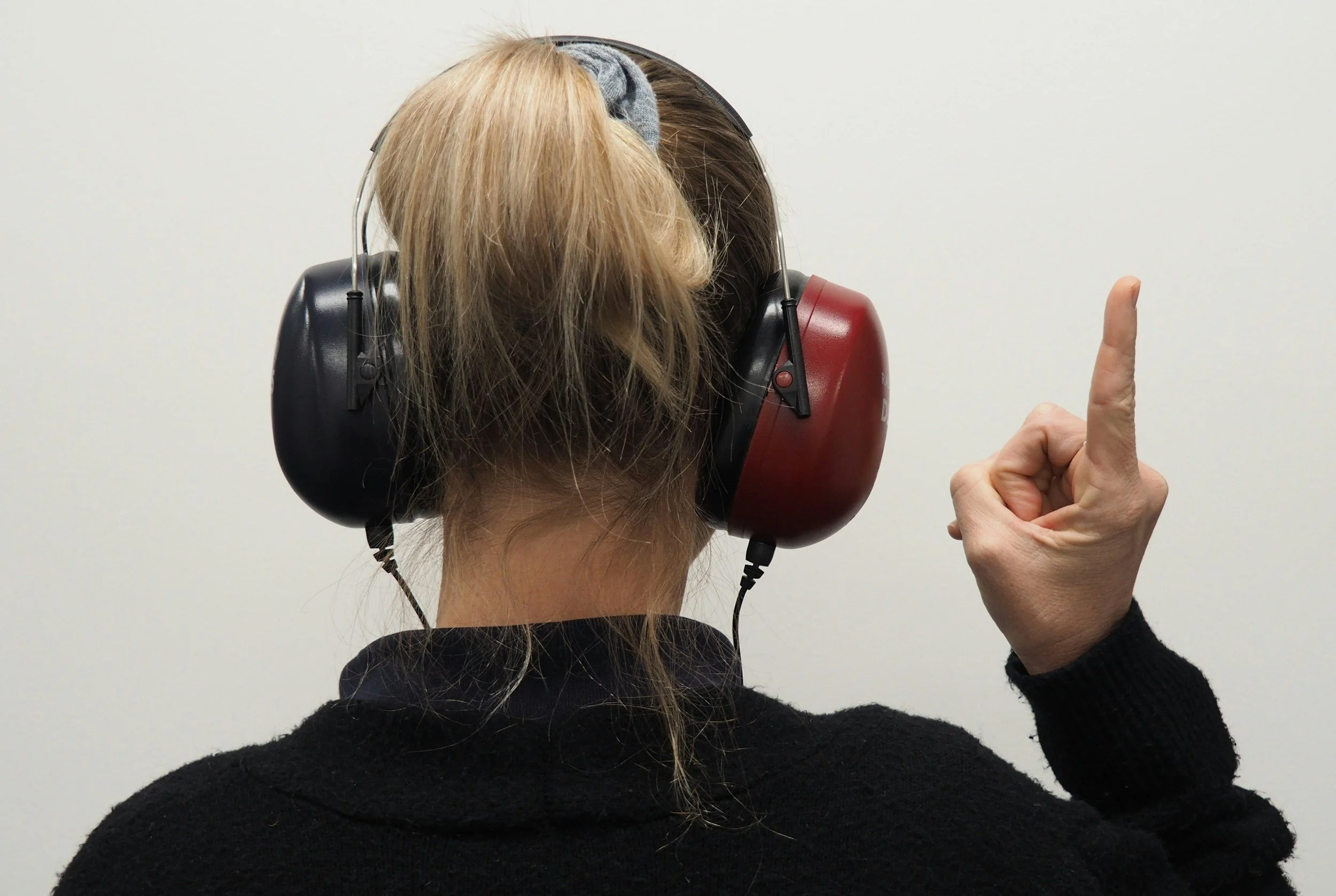
Audiometry and immittance testing are usually done in the same visit, and I ask for them along with the vestibular report. A fluctuating low-frequency sensorineural hearing loss supports a diagnosis of Ménière's disease. A conductive air-bone gap with normal acoustic reflexes suggests a third window disorder. An unexplained asymmetric sensorineural loss may need MRI to rule out a vestibular schwannoma, and that referral comes before rehabilitation decisions.
Normal results
A normal test battery rules out some causes but does not mean the patient has no vestibular problem. These tests measure peripheral reflexes and some central pathways. They are not designed to detect vestibular migraine between attacks, persistent postural-perceptual dizziness, or the visual-vestibular and cognitive-motor integration problems that are common after concussion. A patient with normal lab results and clear functional limitations still needs rehabilitation, and the results help me decide which diagnoses to consider and what to expect from recovery.
When I refer
I refer for vestibular testing when bedside findings are unclear, when I suspect a bilateral loss, when progress stops despite good adherence, when symptoms suggest a third window disorder or otolith involvement, or when the patient reports hearing changes or tinnitus. I include a specific clinical question on the referral, because that gives the audiologist or otolaryngologist a better idea of what to focus on in the report.
References
Halmagyi, G. M., Chen, L., MacDougall, H. G., Weber, K. P., McGarvie, L. A., & Curthoys, I. S. (2017). The video head impulse test. Frontiers in Neurology, 8, Article 258. https://doi.org/10.3389/fneur.2017.00258
McGarvie, L. A., Curthoys, I. S., MacDougall, H. G., & Halmagyi, G. M. (2015). What does the dissociation between the results of video head impulse versus caloric testing reveal about the vestibular dysfunction in Ménière's disease? Acta Oto-Laryngologica, 135(9), 859-865. https://doi.org/10.3109/00016489.2015.1015606
Rosengren, S. M., Colebatch, J. G., Young, A. S., Govender, S., & Welgampola, M. S. (2019). Vestibular evoked myogenic potentials in practice: Methods, pitfalls and clinical applications. Clinical Neurophysiology Practice, 4, 47-68. https://doi.org/10.1016/j.cnp.2019.01.005
Strupp, M., Kim, J.-S., Murofushi, T., Straumann, D., Jen, J. C., Rosengren, S. M., Della Santina, C. C., & Kingma, H. (2017). Bilateral vestibulopathy: Diagnostic criteria. Consensus document of the Classification Committee of the Bárány Society. Journal of Vestibular Research, 27(4), 177-189. https://doi.org/10.3233/VES-170619